Gene therapy is a promising therapeutic approach that has experienced significant growth in recent decades, with gene nanomedicines reaching the clinics.
However, translating more gene nanomedicines from bench to bedside is still challenging. The use of robust preclinical models that can better predict the future behavior of nanosystems is essential to improve their translation process. A suitable option to assess the possibilities of nanomedicine is Zebrafish, which has been proposed as an in vivo platform to evaluate the potential of newly developed nanosystems for gene therapy applications in cancer treatment [1].
Lipid nanosystems are the best delivery approach for gene therapy
One of the main limitations in developing novel gene therapies is the need for efficient carriers capable of protecting and transporting them to their site of action. Viral vectors are the carriers that have moved most quickly to clinical trials, but they accumulate several side effects, such as insertional mutagenesis and high immunogenicity. Non-viral vectors, like lipid nanoparticles and lipid nanoemulsions, have been proven to resolve these limitations successfully.
Cascallar et al. have successfully tested a formulation based on lipid nanosystems and demonstrated its effectiveness for cancer gene therapy in both in vitro and in vivo models [2]. Moreover, it was proposed that Zebrafish embryos can be used as an in vivo model to evaluate their potential even more reliably.
Zebrafish as a highly suitable model for the development of cancer gene therapies
Zebrafish is an attractive in vivo platform that offers several advantages over other in vivo models, such as low maintenance costs, transparency, robustness, and a high homology with the human genome [3].
In nanomedicine, this model is proposed to assess the biocompatibility and toxicity of several nanomaterials and validate their therapeutic efficacy [4]. The presence of organs and metabolic pathways analogous to humans in Zebrafish allows toxicological and biocompatibility evaluations [5].
In the context of cancer, the transparency of embryos and the availability of fluorescently labeled transgenic Zebrafish lines offer the possibility to track cancer cells in xenograft assays or genetic models [6].
On the other hand, the transparency of Zebrafish allows the determination of the toxicity of nanosystems in different anatomical sites and their tracking to establish biodistribution and interaction profiles with tumor cells without the need for invasive techniques [7].
Based on these features, successfully validating the effectiveness of a gene delivery systems in this model represents a promising indicator of its effects on humans.
How lipid nanosystems show their potential in Zebrafish model
The experiments performed by Cascallar and colleagues demonstrated the potential of the developed lipid nanosystems for gene therapy applications, and more specifically, in oncology. Zebrafish enables to observe/visualize the interaction of fluorescent nanoparticles with cancer cells in a more complex system than the one represented by a cell culture since different cell types from the tumor environment are present in the fish.
Gene expression was confirmed after injection in Zebrafish embryos. Biodistribution studies validated the efficacy of the nanosystems to reach disseminated cancer cells. Moreover, the nucleic acids, a plasmid DNA and a microRNA, remained protected and associated to the lipid nanosystems, confirming a high stability in an in vivo environment [2].
DIVERSA aims to grant access to simple and efficient delivery systems
DIVERSA’s R&D efforts have led to the development of pioneering lipid nanosystems to provide the best viral-free alternative to gene therapy applications. DIVERSA’s nanoparticles offer a safe, efficient, non-immunogenic, and biocompatible option for associating innovative and original materials to any type of nucleic acid, like miRNAs and mRNAs.
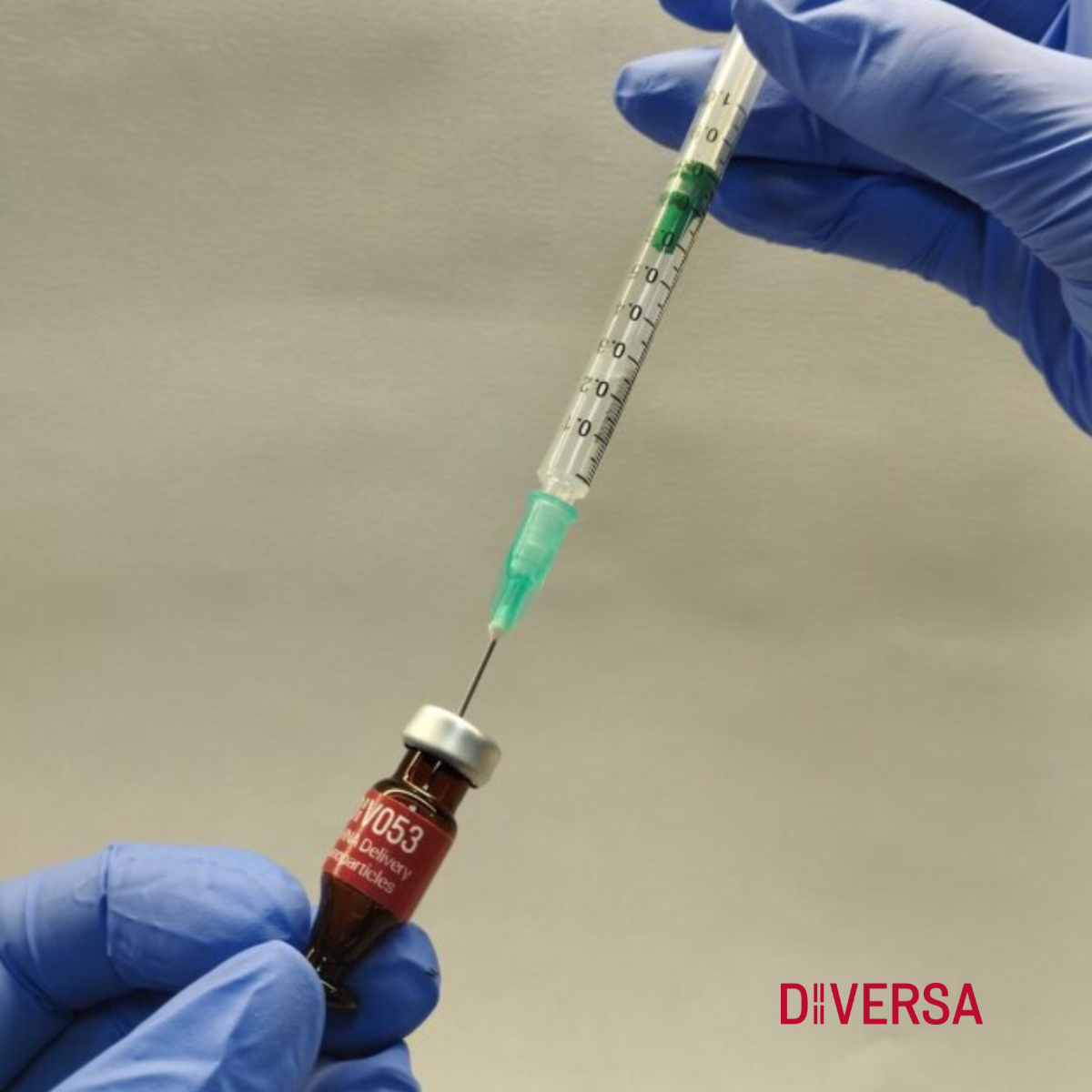
DIVERSA’s new mRNA Delivery Nanoparticles is an innovative formulation that will simplify your mRNA transfection procedures. Our easy-to-use reagent ensures efficient delivery and exceptional transfection performance across various cell types, including in vitro and in vivo models. It requires no special or expensive equipment; a syringe and a micropipette are all you need to formulate and deliver mRNA.
Advancing your gene therapy research towards clinical applications has never been simpler!
Contact us if you require formulations to suit your delivery project! We are looking forward to helping you with your innovative ideas!
References
- Sieber, S.; Grossen, P.; Bussmann, J.; Campbell, F.; Kros, A.; Witzigmann, D.; Huwyler, J. Zebrafish as a Preclinical in Vivo Screening Model for Nanomedicines. Adv Drug Deliv Rev 2019, 151–152, 152–168, doi:10.1016/j.addr.2019.01.001.
- Cascallar, M.; Hurtado, P.; Lores, S.; Pensado-López, A.; Quelle-Regaldie, A.; Sánchez, L.; Piñeiro, R.; de la Fuente, M. Zebrafish as a Platform to Evaluate the Potential of Lipidic Nanoemulsions for Gene Therapy in Cancer. Front Pharmacol 2022, 13, 1007018, doi:10.3389/fphar.2022.1007018.
- Kimmel, C.B.; Ballard, W.W.; Kimmel, S.R.; Ullmann, B.; Schilling, T.F. Stages of Embryonic Development of the Zebrafish. Dev Dyn 1995, 203, 253–310, doi:10.1002/aja.1002030302.
- Cascallar, M.; Alijas, S.; Pensado-López, A.; Vázquez-Ríos, A.J.; Sánchez, L.; Piñeiro, R.; de la Fuente, M. What Zebrafish and Nanotechnology Can Offer for Cancer Treatments in the Age of Personalized Medicine. Cancers (Basel) 2022, 14, 2238, doi:10.3390/cancers14092238.
- Horzmann, K.A.; Freeman, J.L. Making Waves: New Developments in Toxicology With the Zebrafish. Toxicol Sci 2018, 163, 5–12, doi:10.1093/toxsci/kfy044.
- Lawson, N.D.; Weinstein, B.M. In Vivo Imaging of Embryonic Vascular Development Using Transgenic Zebrafish. Dev Biol 2002, 248, 307–318, doi:10.1006/dbio.2002.0711.
- Al-Thani, H.F.; Shurbaji, S.; Yalcin, H.C. Zebrafish as a Model for Anticancer Nanomedicine Studies. Pharmaceuticals (Basel) 2021, 14, 625, doi:10.3390/ph14070625.


